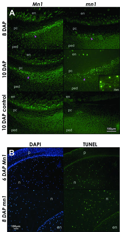
Figure 3.
Apoptotic-like PCD is diagnosed by the TUNEL stain in A, P-C layers of Mn1 and mn1 maize caryopses at 8 and 10 DAP and B, nucellus tissue at 6 and 8 DAP. A, Green fluorescent dots mark nuclei containing fragmented DNA. Magenta arrowheads point to the layers of cells in the P-C region with TUNEL-positive nuclei. The inset in 10-DAP mn1 panel shows ringed foci of faint green fluorescence, indicative of apoptotic like bodies. The 10-DAP control panels, without the TdT enzyme, show only the background green autofluorescence, independent of the TUNEL stain. B, Comparison of DAPI and TUNEL staining in the apical part of the caryopsis. At 6 DAP, the nuclei in the nucellus reacted positively with both DAPI and TUNEL, whereas the pericarp nuclei stained much weaker with TUNEL compared to DAPI. At 8 DAP, the comparison of nucellar and endosperm tissue shows relatively much weaker TUNEL reaction in the endosperm compared to nucellus, indicating that endosperm and pericarp cells did not undergo PCD such as the nucellus cells at this early stage of development (both Mn1 and mn1 genotypes were similar). en, endosperm; n, nucellus; p, pericarp; pc, P-C layer; ped, pedicel.

