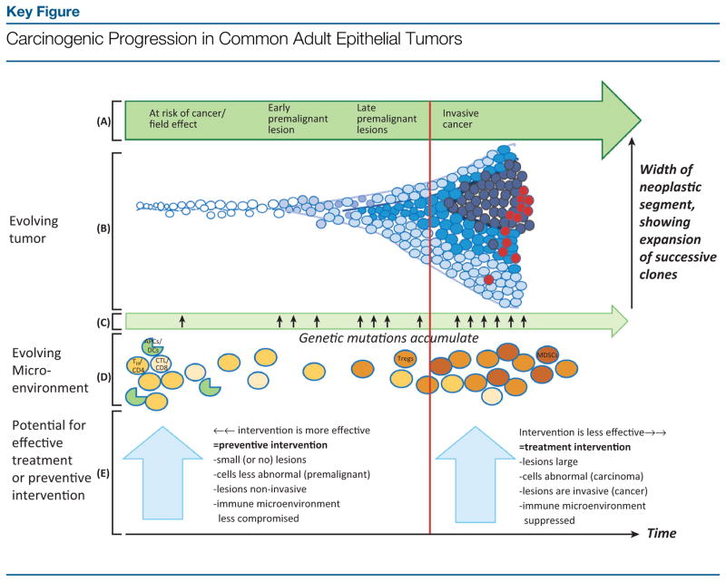
Figure 1.
Evolution of a tumor from normal tissue through progressively advanced premalignant lesions to invasive cancer [going from left to right in (A)] is depicted by the cartoon in (B). Normal cells are shown in white. Progressively abnormal cells appear in gradually darker shades of blue, with dark-blue circles representing invasive cancer cells and red circles depicting cancer cells with metastatic potential. This progression results from the accumulation of oncogenic genetic mutations (C). The resulting mutational accumulation confers growth advantages, leading to clonal expansion over time of genetically more complex cells, as shown in cells spreading out along the Y axis (B). In parallel with the neoplastic progression (B), the microenvironment, in particular the immune environment (D), evolves. In normal-appearing or at-risk tissue, the immune component of the microenvironment is populated in large part by immunocompetent cells capable of fighting cancer cells; in (D), these are represented by TH/CD4 and cytolytic/CD8 T cells and antigen-presenting cells (APCs), such as dendritic cells (DCs). Among others, these cells are capable of carrying out immuno-surveillance and eliminating incipient premalignant cells. As lesions progress to more advanced premalignant and, ultimately, invasive malignant states, the immune environment becomes progressively suppressed and less able to eliminate the abnormal cells. This evolving immunosuppression is represented in (D) by increased abundance of T regulatory cells (Tregs) and myeloid-derived suppressor cells (MDSCs), which antagonize beneficial immune responses, allowing the tumor to grow. Administering anticancer drugs or vaccines before invasion (vertical red line) constitutes ‘preventive’ intervention. This approach, especially early during progression, is likely to be more effective than administering preventive agents late in premalignancy or ‘treatment’ interventions after invasion. The more intact state of the immune system early during premalignancy facilitates a robust response to cancer-preventive vaccination and may also enhance anticancer responses to chemopreventive agents. Additional factors contributing to the relative efficacy of preventive versus treatment interventions are shown in the large light-blue arrows. The progressive changes observed in the tumor microenvironment during carcinogenesis, including evolving immunosuppression, are reviewed in [93,138,139]. Key:
 , normal-appearing/at-risk cell;
 ,
,
 , progressively abnormal premalignant cells;
 , invasive cancer cell;
 , cancer cell with metastatic potential;
 , helper T cell (TH/CD4];
 , cytolytic T cell (CTL/CD8];
 , APC, for example, DC.

