. 2017 Jan 16;6:e20898. doi: 10.7554/eLife.20898

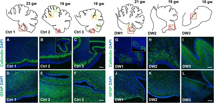
Figure 4. Defects in Purkinje cell alignment and Bergmann glial morphology were observed in the cerebellum of fetuses with chr 6p25 mutations.

(A–L) Midsagittal sections through normal (A-F) and del chr 6p25 (G–L) fetal cerebellar vermis stained for Calbindin (A–C, G–I) and GFAP (D–F, J–L). Cerebellar outlines are provided for clarity. Colored boxes show locations of panels. Ages are indicated in gestational weeks (gw). Purkinje cells during normal cerebellar development were arranged in a distinct multilayered band beneath the molecular layer throughout the cerebellum from 18–23 gw (AC, insets). In all del chr 6p25 cases, Purkinje cells were ectopically broadly distributed in the forming cerebellar cortex (G–I). Bergmann glial fibers extended from the PC layer to the EGL in the normal cerebellum (D–F). These fibers were sparse and highly dysmorphic in the del chr 6p25 cases (J–L). Scale bar = 100 µm.

DOI: http://dx.doi.org/10.7554/eLife.20898.008

Figure 4.

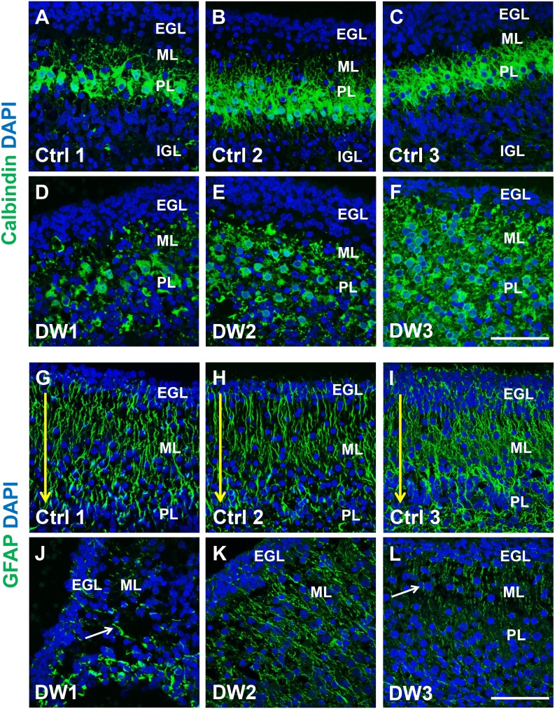
Figure 4—figure supplement 1. Purkinje cell and Bergmann glial fiber morphologies were disrupted in human fetal del chr 6p25 cases.

Figure 4—figure supplement 1.

(A–L) Midsagittal sections through the fetal cerebellar vermis in control fetuses (A–C, G–I) and del chr 6p25 cases (D–F, J–L) stained for Calbindin (A–F; green) and GFAP (G-–L; green). In all control cases, Purkinje cells formed a compact multilayered band beneath the molecular layer, with nascent dendrites projecting into the molecular layer (A–C). In all del chr 6p25 cases, the Purkinje cells were dispersed as a highly disorganized multi-layer zone. Additionally, several cells were ectopically located in the molecular layer (D–F). In control cases, Bergmann glial fibers extended from the EGL to the IGL (G-–I; arrow). There were fewer fibers in all del chr 6p25 cases and their morphology was severely disrupted (J–L, arrows). Scale bar = 50 µm.