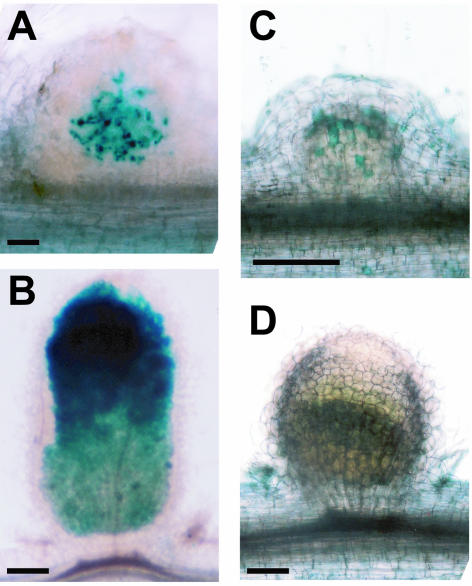
Figure 2.
Rhizobial infection (A and B) and polyphenolic accumulation (C and D) in nip and wild-type nodules. nip and A17 wild-type plants inoculated with S. meliloti carrying the hemA∷lacZ reporter were hand-sectioned and stained with X-Gal at 15 dpi. S. meliloti stain blue. A, nip nodule; B, A17 nodule. nip and A17 wild-type plants were inoculated with S. meliloti, fixed, and stained with potassium permanganate followed by methylene blue. Polyphenolics stain blue in this procedure. C, nip nodule; D, A17 nodule. Bars = 0.2 mm.

