Figure 3.
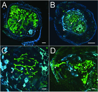
Confocal light micrographs of M. truncatula wild-type (A17) and mutant (nip) nodules. A, Wild-type nodule 13 dpi. Nodule has emerged from the root and shows normal mature infected cells containing intracellular bacteroids (green). A typical infected cell is indicated by the double arrowhead. Bar = 100 μm. B, nip nodule 13 dpi demonstrating abnormal nodule formation. The nodule is infected with bacteria and there are an unusually high number of threads present (single arrowhead). Mutant nodules often did not emerge from the root and autofluorescence (blue) is observed in cells around the infection threads (barbed arrowhead). Bar = 100 μm. C, Higher magnification of nip mutant nodule 13 dpi showing abundant threads that terminate without release of bacteria into plant cell cytoplasm. Threads are thickened, exhibit abnormal bulges (arrowhead), and are flanked by cells exhibiting autofluorescence (blue). Bar = 20 μm. D, High magnification view of nip mutant nodule 29 dpi showing highly enlarged and distorted threads. Single arrowhead indicates an infection thread; barbed arrowhead indicates blue autofluorescence in nodule cells. Bar = 20 μm.

