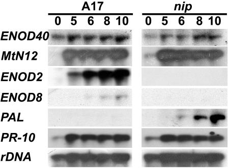
Figure 6.
RNA-blot analysis of gene expression during nip nodule development. Gel blots were prepared from 20 μg of total RNA extracted from A17 or nip roots inoculated with S. meliloti at the indicated times after inoculation (dpi). Blots were hybridized sequentially with radiolabeled ENOD40, MtN12, ENOD2, ENOD8, PAL, and PR-10 probes, with rDNA serving as the loading control.

