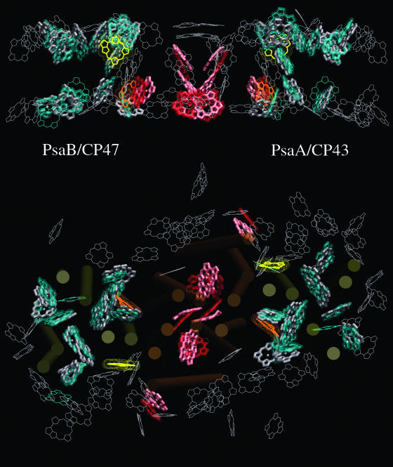
Figure 1.
Comparison of PSI and PSII Structures.
The top panel is a side-on view from within the plane of the thylakoid membrane. The bottom panel is a view from the lumenal side. The individual domains of PSII (CP43, CP47, and D1/D2) are overlapped with the PSI structure as described in the text. Chlorophylls of the RC core domains of PSII (PSI) are shown in red (pink). Antenna chlorophylls of PSII (PSI) are shown in green (gray). Antenna chlorophylls that are conserved in both photosystems are shown with bold lines. Chlorophylls unique to one photosystem are shown with thin lines. The most important optimized connecting chlorophylls of PSII (PSI) are shown in yellow (orange).

