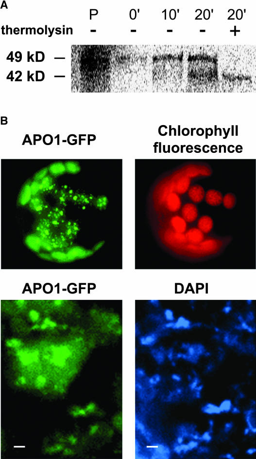
Figure 6.
Sublocalization of APO1 within the Chloroplast.
(A) Import of radiolabeled APO1 proteins into isolated chloroplasts and subsequent detection of gel-separated proteins by phosphor imaging. The precursor of 49 kD (P) was imported and proteolytically processed to 42 kD at the indicated periods of chloroplast incubation. After import chloroplasts were either treated (+) or not treated (−) with thermolysin to digest nonimported proteins.
(B) The APO1 protein was fused to GFP (APO1-GFP) and transiently expressed in tobacco protoplasts. GFP fluorescence was exclusively found in spots inside the chloroplast as revealed by chlorophyll fluorescence (top). Transformed protoplasts were incubated with DAPI and visualized at higher magnification (bottom). Bars = 1 μm.

