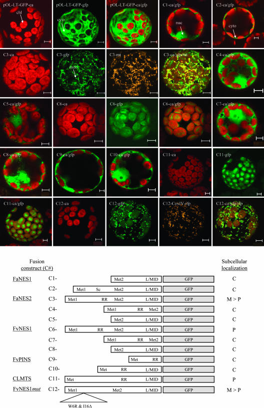
Figure 9.
Transient Expression of GFP Fusions in Tobacco Protoplasts Detected by Confocal Laser Scanning Microscopy.
pOL-LT-GFP is the original vector used to fuse the different strawberry gene fragments to GFP, which directs GFP expression to the cytosol and nucleoplasm (see Methods). Chloroplasts (chl; ∼5 μm in size), mitochondria (mito; ∼1 μm in size), cytosol (cyto), and nucleoplasm (nuc) are indicated by arrows. If several images are shown for a single construct, they are derived from the same protoplast. ca, chlorophyll autofluorescence detected in the red channel; gfp, green fluorescent protein fluorescence detected in the green channel; ca/gfp, combined red and green channels; mt, MitoTracker mitochondrial stain detected in the orange channel or merged with the green and red channels (ca/gfp/mt). Bottom panel, schematic and localization results for each fusion construct. Met1 and Met2, Met residues at the N termini of the proteins (Figure 4); L/MID, conserved motif in terpene synthases (Figure 4); Sc, stop codon; C, cytosol; P, plastids; M > P, more in mitochondria than in plastids. CLMTS, GFP fusion of the 5′ region of a typical monoterpene synthase from Citrus limon (Lucker et al., 2002).

