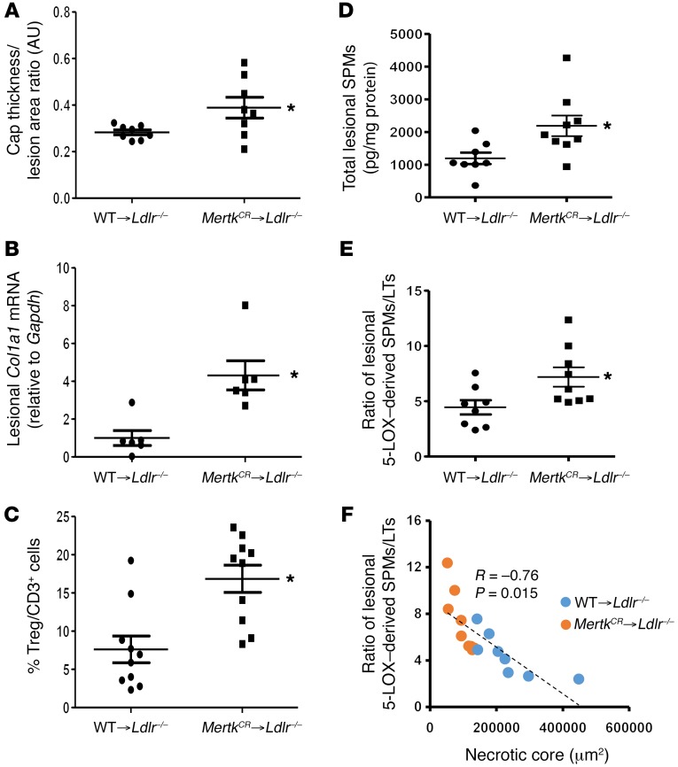
Figure 3. Suppression of MerTK cleavage improves features of resolution in plaques and increases aortic content of specialized proresolving mediators.
(A) Aortic root sections from WT → Ldlr–/– and MertkCR → Ldlr–/– bone marrow–transplanted mice were stained with Picrosirius red. Quantified data are presented as the ratio of fibrous cap thickness to lesion area, expressed as arbitrary units (AU) (n = 8 per group). *P < 0.05. (B) A subset of aortic root sections was chosen randomly for quantification of Col1a1 mRNA by reverse transcriptase quantitative PCR, with normalization to Gapdh mRNA (n = 6 for each group). *P < 0.05. (C) FoxP3+ Tregs and total CD3+ T cells were quantified in aortic root sections by immunofluorescence microscopy and expressed as percentage Tregs/CD3+ cells (n = 10 for each group). The average absolute numbers of these cells, most of which were in the adventitia, were 8 ± 2.49 and 19 ± 3.74 per section for FoxP3+ Tregs (*P < 0.05) and 106 ± 16.13 and 107 ± 14.33 per section for CD3+ cells (NS) for the WT and MertkCR cohorts, respectively. (D and E) Quantification of specialized proresolving mediators (SPMs) and ratio of aortic 5-LOX–derived SPMs/leukotrienes (LTs) in aorta of WT → Ldlr–/– (n = 8) versus MertkCR → Ldlr–/– (n = 9) bone marrow–transplanted mice. *P < 0.05. (F) Correlation of the ratio of 5-LOX–derived SPMs/leukotrienes with necrotic core area (n = 17). P represents the 2-tailed probability value of a Pearson correlation coefficient. A 2-tailed Student’s t test was used for A–E.

