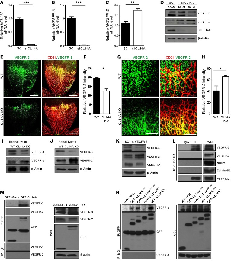
Figure 3. CLEC14A deficiency attenuates VEGFR-3 expression, promotes VEGFR-2 expression, and forms a CLEC14A–VEGFR-3 complex via the CLEC14A cytosolic domain in ECs.
(A–C) Relative GAPDH-normalized mRNA levels of human CLEC14A (hCLEC14A), hVEGFR-3, and hVEGFR-2 after CLEC14A silencing in HUVECs. (D) Reduced VEGFR-3 and increased VEGFR-2 protein expression in HUVECs after silencing of CLEC14A with 50 nM CLEC14A siRNA. (E and F) Confocal images and quantification of relative VEGFR-3 staining intensity in P5 retinae from WT and CLEC14A-KO mice, demonstrating reduced VEGFR-3 in CLEC14A-KO retinae (percentage of control). Scale bars: 500 μm. (G and H) Confocal images and quantification of relative VEGFR-2 intensity of P5 retinae from WT and CLEC14A-KO mice showing increased VEGFR-2 expression (percentage of control). n = 6 per group. Scale bars: 100 μm. (I and J) Decreased VEGFR-3 and increased VEGFR-2 protein expression in retinal and aortal lysates isolated from P5 WT and CLEC14A-KO pups. (K) VEGFR-3, CLEC14A, and VEGFR-2 protein expression in HUVECs after silencing of VEGFR-3 with 50 nM siRNA. VEGFR-3 silencing reduced CLEC14A expression and promoted VEGFR-2 expression. (L) Co-IP of endogenous CLEC14A with VEGFR-3 but not with VEGFR-2 in HUVECs. (M) IP assay showing that overexpressed GFP-tagged CLEC14A (GFP-CL14A) bound to VEGFR-3 but not to VEGFR-2 in HUVECs. Western blot shows transfection of GFP-Mock and GFP-CLEC14A in HUVECs. (N) Clec14a deletion series shows that VEGFR-3 interacted with the cytosolic domain of CLEC14A. IP analysis shows deletion mutants of GFP-tagged CLEC14A (GFP-Mock, GFP-CL14A-ΔLEC, GFP-CL14A-ΔEGF-like, GPF-CL14A-ΔCyto, and GFP-CL14A-FL) interacting with VEGFR-3. The deletion mutants were transfected into HUVECs and immunoprecipitated with anti-GFP antibody or IgG. Western blot analysis of cell lysates after transfection of each deletion mutant into HUVECs. All experiments were repeated at least 4 times with similar results. *P < 0.05, **P < 0.005, and ***P < 0.0001, by paired, 2-tailed Student’s t test. SC, scrambled siRNA; FL, full-length; WB, Western blot; WCL, whole-cell lysate. Error bars represent the mean ± SD.

