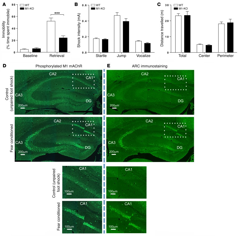
Figure 1. M1 mAChRs play an important role in hippocampal-dependent learning and memory.
(A) Fear-conditioning response of WT and M1-KO mice. Statistical analysis by 2-way ANOVA with Sidak’s multiple comparison test. ***P < 0.001. (B) Pain thresholds for WT and M1-KO mice. Statistical analysis by Student’s t test. (C) Locomotion of WT and M1-KO mice was determined by total distance traveled during an open field test. Data were analyzed using 2-way ANOVA with Sidak’s multiple comparisons. All WT and M1-KO behavioral data are shown as mean ± SEM of n = 8 mice. (D) An antibody-based biosensor for M1 mAChR activation (phosphorylation of the M1 mAChR on S228 in the third intracellular loop) was used to assess M1 mAChR activity in the hippocampus. Following fear-conditioning training, phosphorylation at S228 of the M1 mAChR was increased in the CA1 and CA3 regions and dentate gyrus of the hippocampus relative to control mice that received a 2-second unpaired foot shock. Magnification of the CA1 region (indicated by the rectangle) is shown in lower panels. (E) Fear-conditioning training increased neuronal activity, as assessed by an increase in ARC immunostaining, in the same regions of the hippocampus as those observed for activated M1 mAChR. D and E are composited images. Magnification of the CA1 region (indicated by the rectangle) is shown in lower panels (see D and E). Scale bars: 200 μm (upper panels); 100 μm (lower panels).

