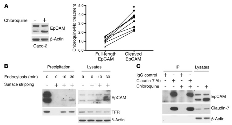
Figure 5. Matriptase cleavage leads to dissociation of EpCAM and claudin-7 and targets EpCAM for internalization and lysosomal degradation.
(A) Caco-2 cells were treated with or without 100 μM chloroquine for 20 hours, and RIPA lysate proteins were resolved using SDS-PAGE and immunoblotted with anti-EpCAM. Band intensities corresponding to full-length EpCAM and matriptase-cleaved EpCAM were quantified and normalized to that corresponding to β-actin intensity in each condition. Paired ratios of band intensities after chloroquine treatment relative to nontreatment controls are depicted (n = 8). The 2-tailed P value (*P < 0.0001) for the comparison of abundances of full-length EpCAM and cleaved EpCAM in chloroquine-treated and untreated cells was determined using a paired t test. (B) Caco-2 cells were labeled with sulfo-NHS-SS-biotin for 30 minutes at 4°C, followed by incubation at 37°C for the indicated times to allow cell surface proteins to be internalized. Cell surface biotin was stripped via treatment with MESNA, cell lysates were prepared, and biotin-labeled proteins were recovered as described in Methods. Proteins were resolved using SDS-PAGE and immunoblotted with anti-EpCAM or anti–transferrin receptor (TFR). Representative data from 1 of 3 experiments are shown. (C) Caco-2 cells treated with or without 100 μM chloroquine for 20 hours were lysed, protein concentrations were normalized, and immunoprecipitations were carried out with anti–claudin-7 Ab or IgG. Immunoprecipitates and lysate proteins were resolved with SDS-PAGE and immunoblotted with anti-EpCAM and anti–claudin-7. Representative data from 1 of 3 experiments are shown.

