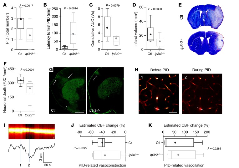
Figure 2. IP3R2-mediated astroglial signaling reduces PID burden without affecting PID-related cerebrovascular changes.
(A–C) Total number of PIDs, latency to first PID, and cumulative AUC as an indicator of overall PID burden were reduced in Ip3r2–/– mice (n = 14) compared with controls (n = 11 mice). (D–G) Infarct volumetry and cell densitometry 72 hours after MCAO revealed a reduced infarct size (n = 15 mice for each group) and a reduced density of dead neurons (quantified by Fluoro-Jade C [FJC]) (n = 15 mice in each group). Arrows in G indicate cortical neurodegeneration. Scale bar: 1 mm (H) Penetrating arterioles showed strong vasoconstriction during PIDs. Left panel shows the vasculature (labeled with Texas Red dextran) during pMCAO before PID; right panel shows the vasculature during PID. Scale bar: 50 μm. (I) Resliced image (indicated by the dashed line in H) used to determine the arteriolar diameter. Scale bar: 5 μm. Spatial measurements of Texas Red fluorescence (lower trace) demonstrate the diameter decrease during the PID. (J and K) The vascular response to PID, consisting of a vasoconstriction followed by a variable vasodilation, remained unchanged in both groups (n = 4 Cx43-ECFP Ip3r2–/– mice; n = 7 control mice). All P values were determined by Mann-Whitney U test.

