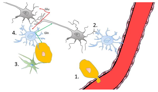
Figure 2.
Brain metastatic cancer cells may exploit a number of unique features (illustrated in 1 to 4) of the brain microenvironment. 1. Brain metastatic cancer cells first need to pass the BBB in order to enter the brain. Once they extravasate, they can benefit from the BBB’s exclusion of large molecules (i.e., therapeutic antibodies) and barring the active efflux of compounds. 2. Astrocytes serve homeostatic roles in supporting neuronal function and survival, but preventing brain colonization of cancer cells by expressing FasL. Cancer cells that evade FasL-mediated killing are able to benefit from astrocytes’ pro-survival functions, rendering brain metastases chemo-resistant. 3. Microglia serve as the resident immune cells of the brain. Microglia have the potential to recognize and eliminate brain metastatic cancer cells by TNF-α and inducible nitric oxide synthase (iNOS). Successful brain metastatic cancer cells must be able to avoid microglial detection. 4. The brain environment serves to support the optimal function of neurons, promoting their survival and preventing excitotoxicity. Glutamate, the essential neurotransmitter, cannot freely exist in the brain parenchyma. Following synaptic glutamate (Glu) release, astrocytes scavenge the excess glutamate and convert it into the non-neuroactive glutamine (Gln), which is then released to be taken up by neurons. Cancer cells may use this glutamine, which exists at relatively high concentrations in the brain, as an important biomolecular building block and energy source. Neurons-gray, astrocytes-blue, microglia-green, endothelial cells-red. Figure drawn in collaboration with Chia-Chi Chang.

