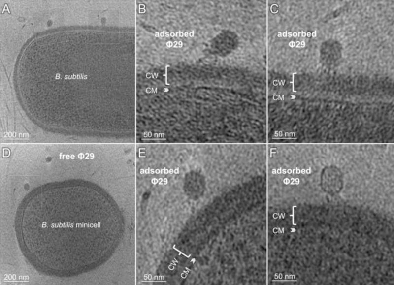
Figure 3. Visualization of B. subtilis infected by Φ29.

A) A central tomographic slice of an infected rod-shaped cell (partial) with diameter ~1μm. B) and C) Zoom-in views highlight two adsorbed phages with different conformations visible in A). CM: cytoplasmic membrane, CW: cell wall. D) A central slice of an infected B. subtilis minicell. E) A zoom-in view from D) highlights an adsorbed Φ29 interacting with the cell wall. F) A separate minicell with an adsorbed Φ29 whose genome has been partially ejected. CW = cell wall; CM = cytoplasmic membrane.
