Fig. 1.
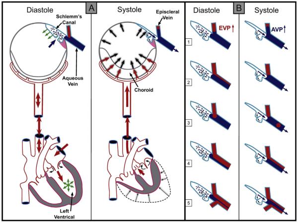
(Panel A-Systole) Cardiac source of pulsatile flow. Systole-induced choroidal vasculature expansion (red arrows). Transient intraocular pressure (IOP) increase (large black arrows). Aqueous pulse wave distends the trabecular meshwork forcing it outward into Schlemm's canal (SC). One-way channels into SC prevent backflow (small curved arrows). Distention of the TM into SC reduces SC volume (black arrows on blue curved surface outlining SC. SC pressure increases. Small black arrow denotes aqueous discharge from SC. Aqueous pulse wave then enters the aqueous vein. (Panel A-Diastole) Blood enters the left ventricle (green circle of arrows). Double red arrows indicate absence of a choroidal pressure wave in diastole. IOP transiently decreases. TM recoils inward during diastole (green arrows). Aqueous enters SC (large blue arrow). (Panel B) During diastole episcleral vein pressure (EVP) is slightly higher than aqueous vein pressure (AVP), resulting in a relative EVP ↑). The EVP ↑ causes episcleral vein blood to move toward or into an aqueous vein. The next systole causes a transient AVP ↑. The oscillations result in pulsatile flow manifestations in the aqueous veins. The AVP ↑ causes transient movement of a standing aqueous wave into a tributary episcleral vein (B1), transient elimination of a lamina of blood (B2), a bolus of blood swept into the increased aqueous stream (B3), an oscillating increase in diameter of the aqueous component of a persistent laminar (B4) or a trilaminar (B5) aqueous flow wave. From: Johnstone M, Jamil A, Martin E. Aqueous Veins and Open Angle Glaucoma. In: Schacknow P, Samples J, editors. The Glaucoma Book. New York: Springer, 2010. p. 65–78.

