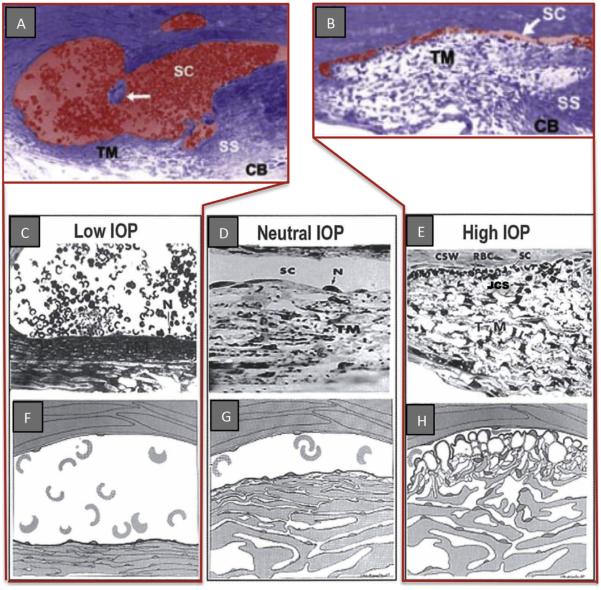
Fig. 3.
Pressure-dependent trabecular meshwork (TM) configuration. A and B are micrographs of two eyes of the same primate with eyes fixed simultaneously in vivo. (A, C, & F); Low IOP; IOP = 0 mm Hg, episcleral venous pressure ~ 8 mm Hg. The scleral spur (SS) is rotated inward toward the anterior chamber. The lumen of Schlemm's canal (SC) is large; the TM tissues are collapsed with obliteration of the juxtacanalicular space (JCS). (D, G) (Neutral lOP) IOP = 0, EVP = 0 the trabecular tissues are in a neutral position, SC lumen size is modest. (B, E, H) (High lOP) lOP = 25 mm Hg, EVP ~ 8 mm Hg during fixation; IOP reduced to 0 mm Hg after fixation with animal still alive. Blood refluxes into SC. The SS is rotated toward SC. The lumen of SC is reduced to a potential space. SC inner wall endothelium distends to reach Schlemm's canal external or corneoscleral wall (CSW). The JCS is large. Large spaces are present between the trabecular lamellae. Red blood cells (RBC) are present in SC but not the JCS. (N, nucleus of Schlemm's canal endothelial cell.) (RBC dimensions ~ 8 μm) From: Johnstone MA. Grant WM: Pressure-dependent changes in structure of the aqueous outflow system in human and monkey eyes, Am J Ophthalmol 75:380, 1973.

