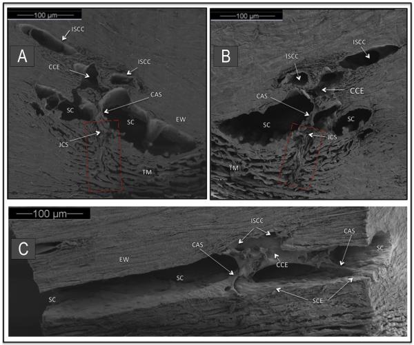
Fig. 4.
Organization of aqueous outflow pathways. Scanning electron microscopy (SEM) of a macaca nemestrina monkey eye. (A, B) are paired mirror image radial sections of trabecular meshwork (TM), Schlemm's canal (SC) and collector channel ostia (CCO). A cylindrical attachment structure (CAS) has a lumen that communicates with the juxtacanalicular space (JCS) and the collector channel entrance (CCE). Red rectangle indicates TM region organized parallel to probable path of preferential aqueous flow into large open funnel at origin of a CAS. Intrascleral collector channel (ISCC). SC external wall (EW). (C) SEM section parallel to the circumference of SC. CAS span across SC to hinged flaps at CCE. A direct path for aqueous flow from SC into the CCE and ISCC is visible.

