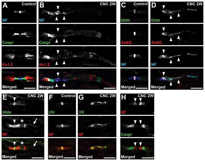
FIGURE 2.
Disrupted molecular organization at nodes. (A,B) Sections of control (A) or compressed nerves (CNC, 2 weeks) (B) are immunostained as indicated. (B) Neurofascin (NF) and Caspr staining on the right side of the nodes are remarkably reduced and dispersed. Arrowheads indicate disrupted clusters of NF. (C,D) Sections of control (C) or compressed nerves (2 weeks) (D) are immunostained for gliomedin (Gldn) in green, ankyrinG (AnkG) in red, and NF in blue. (D) Gliomedin and NF are partially co-localized with disorganized nodal ankyrinG clusters (arrowheads). (E) Section of compressed nerve (2 weeks) is immunostained for gliomedin in green and NF in red. The paranode on the right side of the node is disrupted (asterisk), whereas the gliomedin cluster is preserved (arrowhead). Arrow indicates NF155 enriched at inner mesaxon of the myelin. (F,G) Sections of control (F) or compressed nerves (2 weeks) (G) are immunostained for βIV spectrin (βIV) in green, and NF in red. (G) Clusters of both βIV spectrin and NF are disorganized. (H) Section of compressed nerve (2 weeks) is immunostained for NF in red and Caspr in green. Arrowheads indicate heminodal NF clusters that are present very close. Scale bars = 10 μm (A–H).

