FIGURE 3.
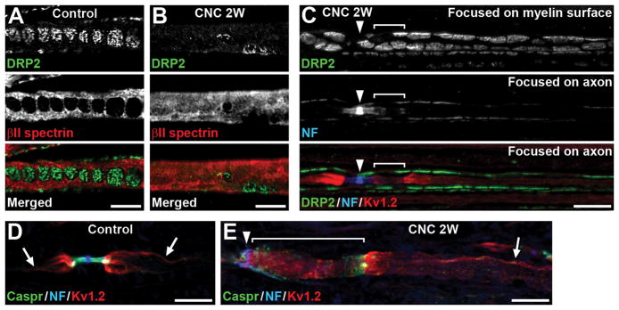
Paranodal disruption can occur without myelin damage in internodes. (A,B) Teased fibers of control (A) or compressed nerves (CNC, 2 weeks) (B) are immunostained for dystrophin-related protein 2 (DRP2) (green) and βII spectrin (red). (B) DRP2 immunostaining is remarkably reduced, and βII spectrin staining is diffusely present. (C) Teased fiber of compressed nerve (2 weeks) is immunostained for DRP2 (green), NF (blue), and Kv1.2 (red). Arrowhead indicates node of Ranvier. Top panel shows DRP2 staining focused on the outer surface of myelin sheath. Middle and bottom panels are focused on the axon. NF staining is dispersed and elongated at the paranode on the right side of the node with partial overlap of Kv1.2 staining (bracket). Note the characteristic cobble stone-like pattern of DRP2 staining is preserved. (D,E) Sections of control (D) or compressed nerves (2 weeks) (E) are immunostained for Caspr (green), NF (blue), and Kv1.2 (red). Arrows indicate Kv1.2 enriched at the axolemma where it apposes the inner mesaxon of the myelin sheath. (E) Kv1.2 staining in internode is preserved (arrow), whereas paranode is remarkably elongated and disorganized with Kv1.2 mislocalization (bracket). Arrowhead indicates node of Ranvier. Scale bars = 10 μm (A–E).

