FIGURE 5.
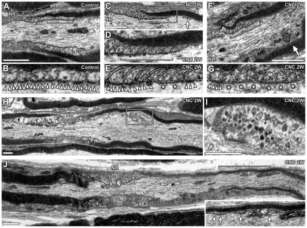
Disrupted paranodal fine structures. TEM images of longitudinal sections of compressed nerves (2 weeks). (A) Normal paranode. (B) Enlarged image of the designated area in panel A (bracket) showing transverse bands (arrowheads). (C) Paranode in compressed nerve. Arrow indicates remote lateral loop. (D) Enlarged image of the boxed area in panel C. Myelin lamellae and lateral loops are preserved. (E) Enlarged image of lateral loops at the designated area in panel D (bracket). Transverse bands (arrowheads) are absent in two lateral loops (asterisks). (F) Paranode in compressed nerve. Arrow indicates enlarged axon with abnormal mitochondria accumulation. (G) Enlarged image of lateral loops in the designated area of panel F (bracket). Transverse bands (arrowheads) are absent in 6 innermost lateral loops (asterisks). (H) Focal axon enlargement in compressed nerve (bracket). (I) Enlarged image of the boxed area in panel H. Abnormal mitochondria accumulation. (J) Myelinated axon in compressed nerve. Paranode on the right side of the node is remarkably disorganized. Inset is an enlarged image of designated area (bracket) showing remote lateral loops (arrows). Myelin sheath on the right side is thinner than the left side. NR indicates location of node of Ranvier (A,C,F,H,J). Scale bars = 1 μm (A,C,F,H,I,J).

