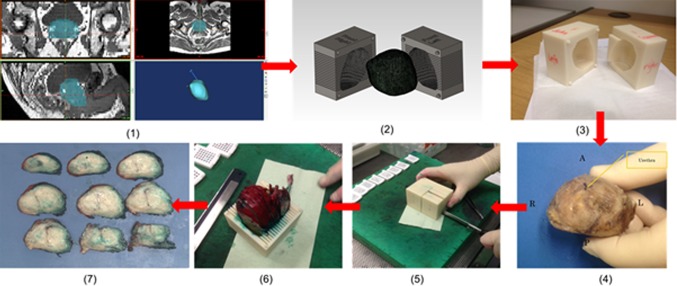
Fig. 2.
Steps of patient-specific molds fabrication and histopathological sectioning—1 segmentation of MRI data in biomedical software MIMICS, 2 mold fabrication in CAD software SolidWorks, 3 3D printout from rapid prototyping machine MakerBot, 4 post-radical prostatectomy specimen before dyeing and mold placement, 5 slicing of prostate specimen with a single blade, 6 sliced sections shown in the mold and 7 specimen slices arranged from apex to base

