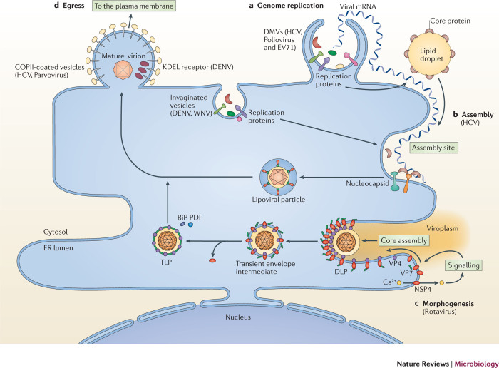
Figure 3. Co-opting the ER to promote the later stages of infection.
Viruses can co-opt functions that are associated with the endoplasmic reticulum (ER) to achieve the four crucial later steps in infection — replication, assembly, morphogenesis and egress. a | Genome replication. During this process, the replication proteins of numerous viruses rearrange the ER membrane to generate membranous structures with different morphologies and terminologies, such as invaginated vesicles (for dengue virus (DENV) and West Nile virus (WNV)) and double-membrane vesicles (DMVs; for hepatitis C virus (HCV), poliovirus and enterovirus 71 (EV71)). These replication sites act to increase the local concentrations of viral and host components that are essential for RNA replication, and enable different steps of the replication process to be coordinated efficiently. b | Assembly. Virus assembly can be tightly coupled to, and coordinated with, genome replication, as exemplified in HCV. To initiate the assembly of viral progeny, a lipid droplet recruits viral core proteins to its surface and delivers them to the site of assembly. In one model of HCV virion assembly, core proteins capture the newly replicated positive-sense RNA ((+)RNA) that extrudes from the neighbouring DMV, forming the nucleocapsid. The nucleocapsid may then bud into the lumen of the ER, generating a newly assembled enveloped virus particle that contains the structural glycoproteins E1 and E2. c | Morphogenesis. Morphogenesis of the assembled HCV particle continues in the ER, with the acquisition of lipoproteins on its surface to generate the mature 'lipoviral' HCV particle that is poised to exit the host cell. For rotavirus, ER-dependent morphogenesis is initiated when its membrane protein non-structural protein 4 (NSP4) induces calcium release from the ER. This triggers a signalling cascade that delivers the structural proteins VP4 and VP7, with assistance from NSP4, to the ER membrane assembly site. The VP4–NSP4–VP7 complex recruits the double-layer particle (DLP) and deforms the membrane to form a transient enveloped intermediate in the lumen of the ER. Following the removal of the ER-derived lipid bilayer, VP7 correctly assembles on the surface of the mature infectious triple-layer particle (TLP), with the simultaneous release of NSP4. The morphologically matured virion then exits the host cell through lysis or secretion. d | Egress. The final step of infection is egress of the mature virion. Viruses that mature in the ER co-opt the ER-dependent secretory pathway to access the extracellular milieu. Examples of using this strategy can be found in egress of the mature HCV, rotavirus and parvovirus particles. Additionally, the ER could also have specific components, such as the KDEL receptor in the case of DENV, that are hijacked to promote exit. COPII, coat protein complex II.

