Fig. 3.

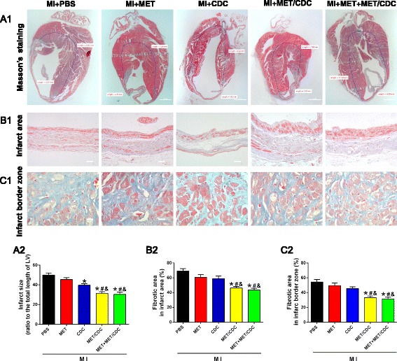
Combination of metformin (MET) treatment and cardiosphere-derived cell (CDC) transplantation reduced infarct size in myocardial infarction (MI) mice. a1 Representative images of Masson’s trichrome staining for heart tissue obtained from hearts with different treatments at 4 weeks post-MI. Scale bar = 1 mm. a2 Graphic representation of the left ventricular (LV) infarct size calculated as the ratio of midline length of the infarcted LV wall to the midline length of total LV wall (n = 5). b Representative images (b1) and quantification (b2) of the fibrotic area in the infarct area 4 weeks post-MI. Scale bar = 200 μm. c Representative images (c1) and quantification (c2) of the fibrotic area at the infarct border zone 4 weeks post-MI. Scale bar = 100 μm. n = 5. Data were analyzed by one-way ANOVA with post-hoc comparisons by the Tukey’s test. *P < 0.05 vs. MI + phosphate-buffered saline (PBS); # P < 0.05 vs. MI + MET; &P < 0.05 vs. MI + CDC. MET/CDC MET-pretreated CDC
