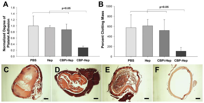
Figure 5.
(A) Platelet adhesion to arterial ECM treated with PBS, heparin sodium, CBPi–heparin, or CBP–heparin (n = 3). The number of platelets binding to ECM treated with each condition was normalized to the number of platelets adherent to ECM treated with PBS alone as a reference. (B) Degree of clot formation by recalcified whole blood on arterial ECM with each separate modification (n = 3). H&E images show the degree of blood clot formation on ECM treated with (C) PBS, (D) heparin sodium, (E) CBPi–heparin, or (F) CBP–heparin. Percent clot formation was determined by comparing mass of the blood clot formed to the mass of the ECM graft before addition of whole blood. Scale bar = 200 μm.

