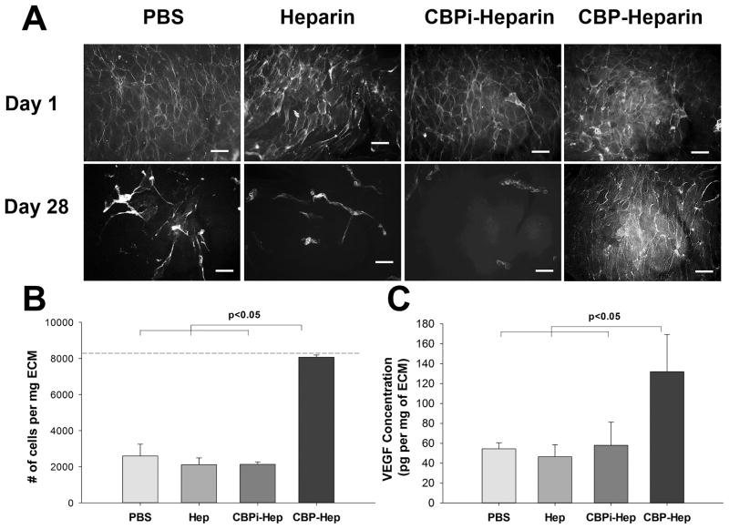
Figure 6.
(A) Phalloidin staining of endothelial cells seeded on the lumen of arterial ECM treated with PBS, heparin sodium, CBPi–heparin, or CBP–heparin at days 1 and 28 (scale bar = 100 μm). (B) Quantitative analysis of adherent cell number on ECM at day 28, with the dash line indicating the initial cell seeding density at day 1 (n = 3). (C) Quantitative analysis of VEGF concentration on ECM after incubating in endothelial cell complete medium (EGM-2, n = 3).

