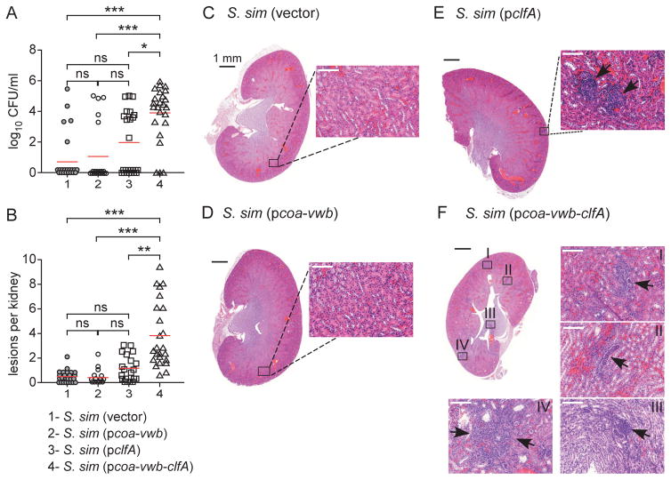
Fig. 6.
Pathogenic conversion of S. simulans with S. aureus coa, vwb and clfA. Mice were injected in the peri-orbital venous plexus with 5×10⁷ of S. simulans carrying either vector, pcoa-vwb, pclfA or pcoa-vwb-clfA, and euthanized 5 days following challenge. (A) Following necropsy, renal tissues were assessed for bacterial load. (B) Lesions observed in panels C through F were enumerated using 20–25 slides per infected group. The data in panels A and B represent the cumulative result of three independent experiments and were analyzed using the one-way ANOVA on ranks (Kruskal-Wallis test) with Dunn's post-hoc test (***, P < 0.001; **, P < 0.01; *, P < 0.05; ns, not significant). (C-F) Microscopy images of hematoxylin-eosin-stained, thin-sectioned renal tissue. Whole kidney sections are shown and areas boxed in black are shown as a 20× enlarged images. Images and insets in panels C and D did not reveal pathological features in renal tissues. In panels E and F, arrows point to replicating bacteria and immune cell infiltrates.

