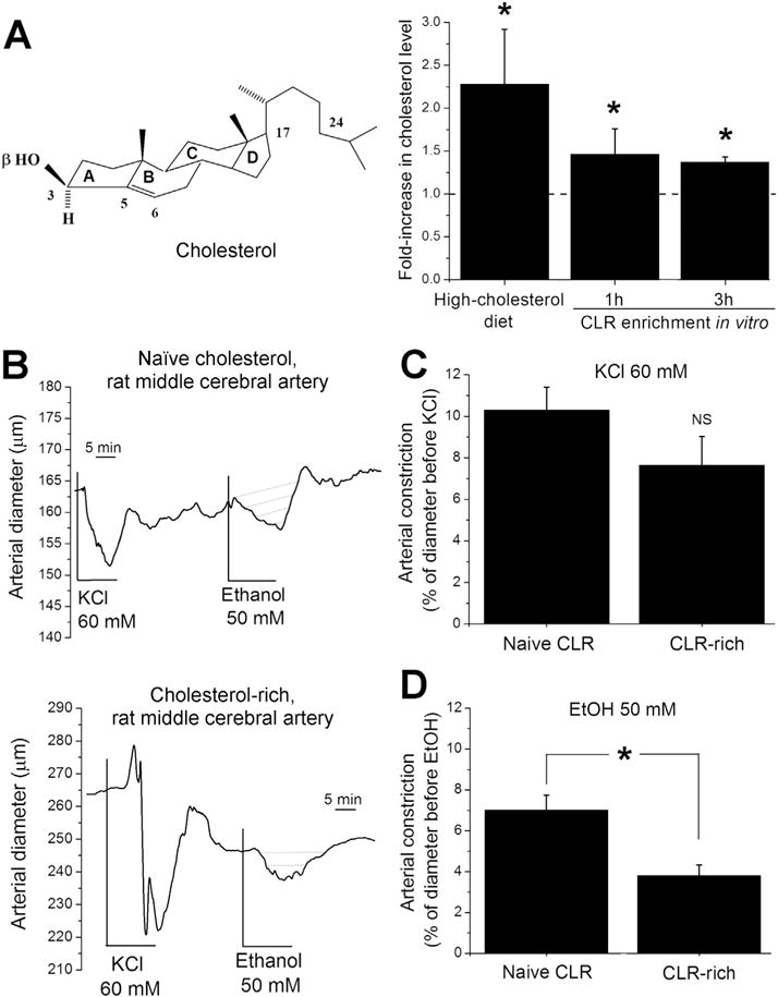
Fig. 1.

In vitro cholesterol enrichment of rat middle cerebral arteries blunted ethanol-induced constriction. (A) Averaged data showing similar fold-increase in artery cholesterol level observed in cerebral arteries from rats on a high-cholesterol diet (18–23 weeks of diet, n = 5) when compared to in vitro cholesterol enrichment (1 h and 3 h following the in vitro cholesterol-enriching treatment, n = 3). *Different from naïve cholesterol (P < 0.05). A dashed line indicates no change in cholesterol level. The insert shows the chemical structure of cholesterol. (B) Original traces showing changes in middle cerebral artery diameter in response to 60 mM KCl, 50 mM ethanol, and Ca2+-free solution in arteries with naïve cholesterol levels vs. cholesterol-enriched vessels. (C) Averaged constriction by 60 mM KCl (n ≥ 10 in each group). (D) Averaged constriction by 50 mM ethanol (n ≥ 6 in each group). *Different from naïve cholesterol (P < 0.05).
