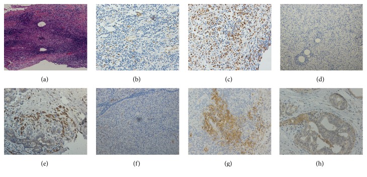
Figure 2.
Representative illustrations of the expression of cytokines in PDM and normal breast tissues. (a) Low-power magnification of PDM (hematoxylin and eosin, ×40). (b) Low expression of IFN-γ (IHC, ×200). (c) High expression of IFN-γ (IHC, ×200). (d) Low expression of IL-12A (IHC, ×100). (e) High expression of IFN-γ (IHC, ×200). (f) Low expression of IL-17A (IHC, ×100). (g) High expression of IL-17A (IHC, ×200). (h) The expression of inflammatory cytokines in normal breast ductal epithelium and stromal cells (IHC, ×200).

