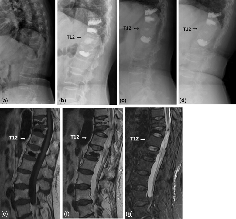
Figure 5.
Images obtained in a 63-year-old woman with OVCFs. (a) The lumbar lateral radiograph before procedure. (b) T10, T11 and L2 were treated with vertebral cement augmentation, where T12 was a sandwich vertebra. (c) Follow-up after 12 months showed no loss of T12 height. (d) Follow-up after 22 months showed no loss of T12 height. (e and f) T1- and T2-weighted images indicated low intense signal, indicating the calcium phosphate component which remains intact for years providing an osteoconductive matrix for new bone ingrowth. (g) Image showed no high signal intensity

