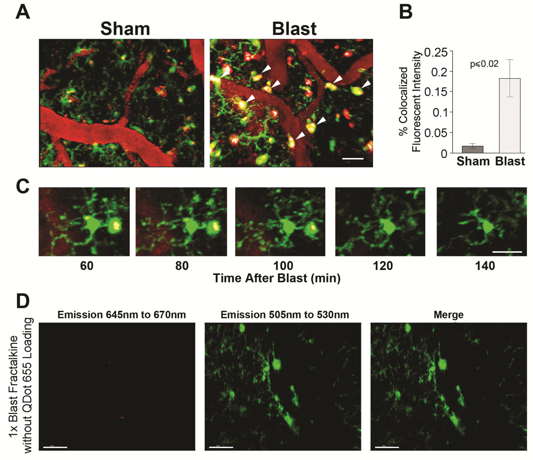
Figure 3. Blast-exposed microglia accumulate Qdots in cell bodies and phagosome-like juxtavascular processes.
(A) In CX3CR1-GFP+/− mice, QDots655 (red) remained predominantly in microvessels in sham-treated mice. In 1X blast-exposured animals, QDots655 crossing the endothelium accumulated in juxtavascular processes and somas of microglia/macrophages (green). QDots655 colocalized with microglia/macrophages denoted by arrowheads. Other red puncta not colocalizing with GFP-positive cells is likely indicative of damaged/fragmented cells. (B) Mild blast caused a significant increase in Qdots655 internalized in microglia/macrophages compared to sham treated animals (p< 0.02). (C) Time lapse image series (60–140 min post-blast) show phagocytosed QDots655 accumulating in enlarged perivascular processes extending from a microglial cell body. (D) Lack of QDots655 emissions (645 nm to 670 nm) in CX3CR1-GFP+/− mice (emissions at 505nm to 530nm) without QDots655 confirms that QDot colocalization in GFP-expressing cells is specific and not due to non-specific auto-flourescence. Scale bars: (A, C) 20 μm, (D) 30 μm.

