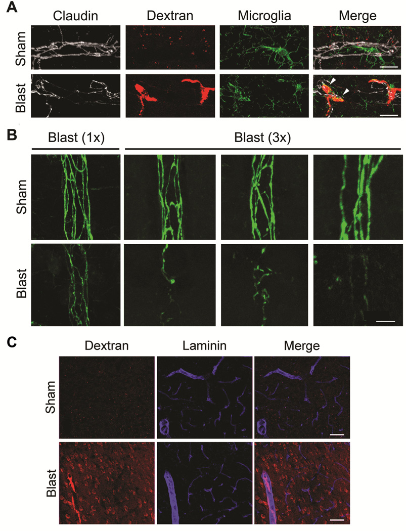
Figure 4. Discontinuous tight junction morphology and endothelial disruption induced by mild blast.
All images show maximum-field projections of 40 serially acquired images scanned at 1 μm intervals in the Z-plane (40 μm total tissue thickness) in cortex of fixed, immunostained sections from wild-type C57BL6 mice. (A) Triple-label confocal microscopy shows tight junctional claudin-5 (white pseudo color) and microglial/macrophage Iba-1 (green) immunoreactivity, as well as 10 kDa dextran (red) that had crossed the BBB one hour post blast. In contrast to sham (upper panel), 1X blast exposure caused focal vascular disruption evidenced by escape of 10 kDa dextran into the surrounding paranchyma (arrowheads) associated with aberrant tight junction claudin-5 morphology and activated-appearing microglia/macrophages. (B) Claudin-5 immunostaining in sham-treated animals (24 hours post-treatment) revealed normal appearing tight junction morphology in cortical penetrating vessels. At 24 hours after a single (1X) blast exposure, disturbed tight junction morphology was markedly less pronouced than at 4 hours after 1X blast-exposure as in Panel A. However, following repetitive 3X blast exposure claudin-5 immunostained tight junctions appear discontinuous and irregular compared to shams. (C) Even though 1X blast induced transiently disturbed tight junction morphology, within 1 hour peripherally-injected 10kDa dextran escapes into the cortical parenchyma (images representative of 3/4 blast-exposed animals and 4/4 shams). Scale bars: (A) 20 μm, (B) 10 μm. (C) 40 μm.

