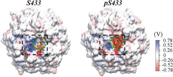
Figure 3.

Electrostatic potential for the initial structure of KLHL3 Kelch domain with unphosphorylated (left) or phosphorylated S433 (right). The surface electrostatic potential for the acidic motif‐binding site (in black dashed rectangle) and that for S433/pS433 (in the green dashed circle) are shown. The initial structure of acidic motif (in yellow) is also indicated.
