Figure 5.

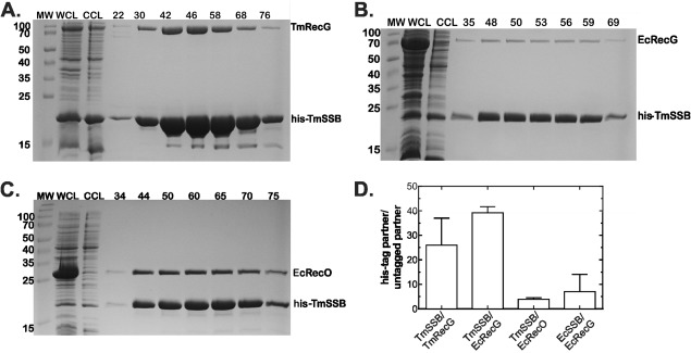
TmSSB forms stable complexes with TmRecG and EcRecO but not EcRecG. The 1‐L cultures of cells expressing RecG (Ec or Tm) with his‐TmSSB were grown to early log phase, IPTG added to 500 µM and growth continued until early stationary phase. Cells were harvested by centrifugation, lysed and the cleared cell lysate applied to a 5‐mL nickel column as described in the Materials and methods. Proteins were then eluted using an imidazole gradient following extensive washing to remove unbound proteins. Aliquots from various fractions throughout the purification were subjected to electrophoresis. Coomassie stained, 15% SDS‐PAGE gels showing various stages during the purification are presented. (A) TmRecG binds to TmSSB. (B) EcRecG does not bind appreciably to TmSSB. (C) TmSSB binds to EcRecO. (D), Analysis of the gels in panels A–C. The data for EcSSB/EcRecG are from column 1, Figure 3(D) and are presented here for comparison. MW, molecular weight marker; WCL, whole cell lysate; CCL, cleared cell lysate; numbers indicate fraction numbers from the peak elution profile.
