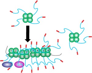
Figure 8.

SSB linkers mediate all protein–protein interactions. Schematics of wild type SSB proteins binding to ssDNA and to partners. The colouring of SSB monomers is as follows the core domain in green, linker in blue and acidic tip in red. Here tetramers have their functional C‐terminal domains exposed in solution. Upon binding to ssDNA the IDLs of monomers 1 and 2, bind to the OB‐folds of monomers 1′ and 2′, respectively. Concurrently, the linkers of monomers 1′ and 2′ bind to the OB‐folds of monomers 3 and 4, and their IDLs bind to monomers 3′and 4′, respectively. The C‐termini of subunits 3′ and 4′ are available to bind to an incoming tetramer as shown on the right. On the opposite side of each tetramer, C‐termini are available for binding to interactome partners such as RecG or RecO (purple and blue ovals). For simplicity, SSB–SSB interactions are shown in the top subunits only, and SSB interactome partners binding in the lower subunits.
