
Figure 2.
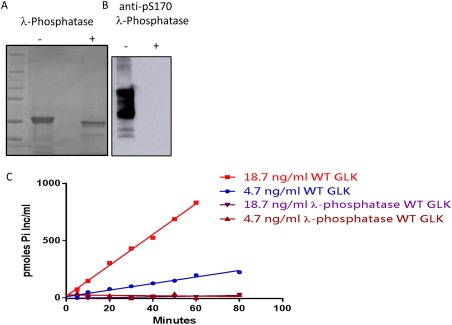
Phosphorylation of WT GLK impacts its activity. (A) Coomassie stained SDS‐PAGE of WT GLK before and after dephosphorylation with lambda‐phosphatase. (B) Phosphorylation state of WT GLK at S170A as measured by western blot with an anti‐S170 phospho antibody. (C) Phosphotransfer activity of increasing amounts of WT GLK over time (red and blue) compared to dephosphorylated WT GLK (brown and purple). The dephosphorylated protein has no measurable activity over the time range of 80 min.