
Figure 3.
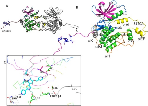
Overall structure of GLK S170A bound to compound 1. (A) Structure of the crystallographic dimer. (B) Close‐up of the GLK S170A monomer demonstrating the N‐terminal strands (purple) the C‐terminal lobe (mainly green helices), the last structured helix (gray; α−K), the ordered C‐terminus (red), and the activation loop (yellow). Ala170 is located between the α‐AL and the alpha EF region. The highly acidic region of the c‐terminal extension is highlighted in blue sticks. (C) Structure of the active site demonstrating residues in close proximity to compound 1. H‐bond networks that demonstrate the active conformation of this kinase are highlighted through sticks (K45‐E61) and in the catalytic loop (D136, K138—neighbor T174). An interactive view is available in the electronic version of the article.