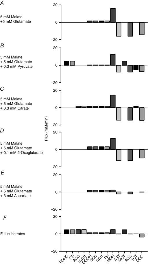
Figure 8. Activities of representative enzymes and substrate transporters at different combinations of substrates in the simple cardiac cell model .

A, 5 mm malate and 5 mm glutamate. B, 5 mm malate, 5 mm glutamate and 0.3 mm pyruvate. C, 5 mm malate, 5 mm glutamate and 0.3 mm citrate. D, 5 mm malate, 5 mm glutamate and 0.1 mm 2‐oxoglutarate. E, 5 mm malate, 5 mm glutamate and 3 mm aspartate. F, full substrates; 1 mm malate, 5 mm glutamate, 0.3 mm pyruvate, 0.3 mm citrate, 0.1 mm 2‐oxoglutarate and 3 mm aspartate. All the simulations were done at 0.3 μm [Ca2+]cyt and 1.5 × 10⁻⁴ mm ms−1 k ATPuse ( = 8.8 mm min−1 except for E, 3.2 mm min−1).
