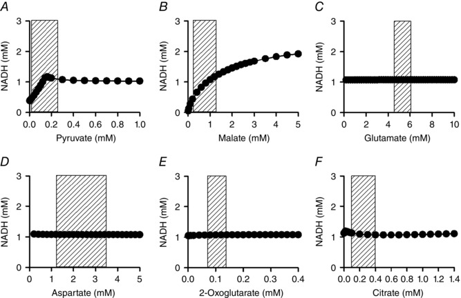
Figure 11. Substrate dependence of NADH in the simple cardiac cell model .

The basic combination of substrates was the same as in Fig. 9. Pyruvate (A), malate (B), glutamate (C), aspartate (D), 2‐oxoglutarate (E) and citrate (F) were systematically increased in the presence of 0.3 μm [Ca2+]cyt. k ATPuse was set to 1.5 × 10⁻⁴ mm ms−1 ( = 8.6–8.9 mm min−1). Shaded areas show physiological range of each substrate (Albe et al. 1990; Kato et al. 2010).
