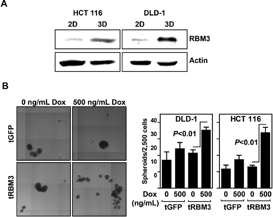
Figure 4.
RBM3 overexpression increases spheroid formation. (A) Western blot analyses for RBM3 from HCT 116 and DLD-1 cells grown in regular tissue culture dish (2D culture) and ultra-low attachment plates (3D culture). Data shows that cells grown in 3D cultures have higher levels of RBM3 compared to 2D cultures. (B) Representative composite image of HCT 116 GFP or RBM3 inducible cells induced for 72 h with 0–500 ng/mL of Dox then plated in ultra-low attachment plates with colonosphere media and allowed to form spheroids for 14 d. Quantification of HCT 116 and DLD-1 spheroids was performed by Celligo embryoid body counting system. Bar graph shows spheroid number from 2500 cells.

