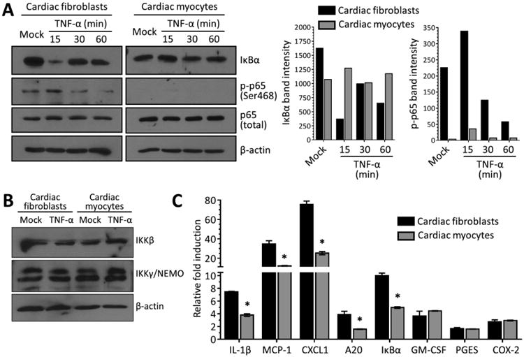
Figure 1. The magnitude of NF-κB activation after TNF-α stimulation is cardiac cell type-specific.

(A) Primary cardiac myocyte or cardiac fibroblast were stimulated with 50 ng/ml TNF-α or media (‘mock’) and whole-cell protein lysates were harvested at the indicated times post-stimulation. Protein lysates were resolved by SDS-PAGE, transferred to a nitrocellulose membrane, and immunoblotted using the indicated antibodies. (B) Protein lysates from primary cultures stimulated for 1 h with media or TNF-α were resolved and transferred as in (A) and immunoblotted using the indicated antibodies. (C) Primary cultures were stimulated with TNF-α as in panel A for 60 min prior to mRNA harvest. Levels of mRNA expression for the indicated genes was analyzed by qRT-PCR and normalized to GAPDH expression. Fold induction by TNF-α is expressed relative to ‘mock’ for each culture (mean ± SD) for a representative of at least two independent experiments. *, Significantly different from cardiac fibroblasts (P < 0.05).
