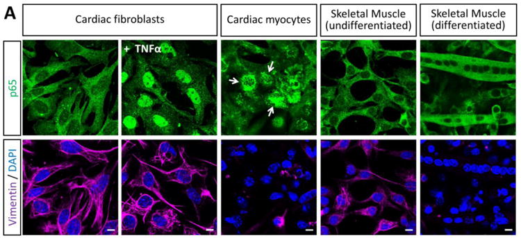
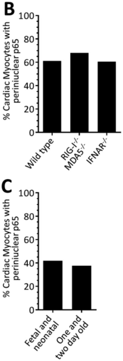
Figure 2. NF-κB localizes to perinuclear compartments in cardiac myocytes.


(A) Primary cardiac myocyte, cardiac fibroblast, or skeletal muscle cultures were fixed for immunofluorescent microscopy using antibodies against p65 and vimentin. Nuclei were counterstained with DAPI. Scale bar = 20 μm. (B) Primary cardiac myocyte cultures generated from the indicated mouse strain were immunostained as in panel A and the percentage of cardiac myocytes (defined as ‘vimentin negative’) were scored according to the localization of p65 in each cardiac myocyte (n = 104 – 214 per case) for a representative experiment. (C) Cardiac myocyte cultures were generated from either ‘fetal and neonatal’ or ‘one- and two-day-old’ wild-type mice and scored as in panel B.
