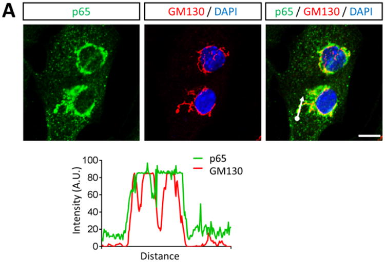
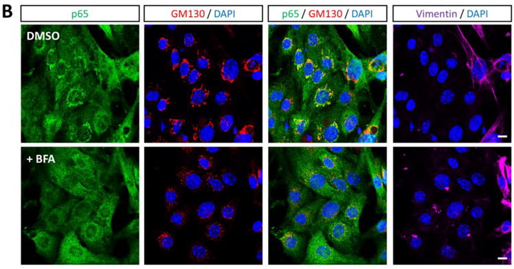
Figure 3. NF-κB perinuclear compartments are associated with the cis-Golgi.


(A) Primary cardiac myocytes were fixed for immunofluorescent microscopy using antibodies against p65 and GM130. Nuclei were counterstained with DAPI. Histograms display measured fluorescence intensity along the drawn line in the overlay panel. (B) Cardiac myocytes were treated with DMSO (vehicle) or 10 μg/ml BFA for 6 h. Cells were then fixed and immunostained as in panel A in addition to vimentin. Scale bar = 10 μm
