
Figure 7. Virus-induced NF-κB activation is cardiac cell type-specific.
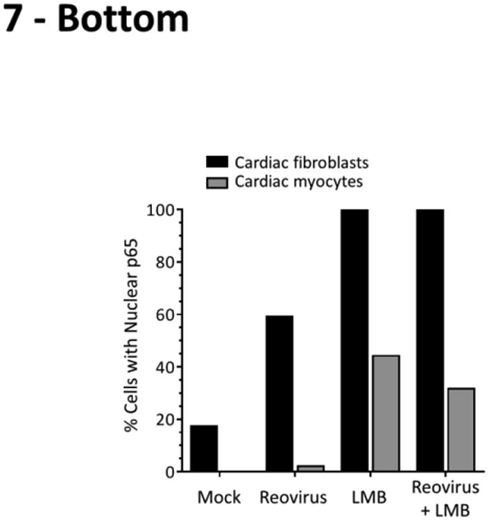
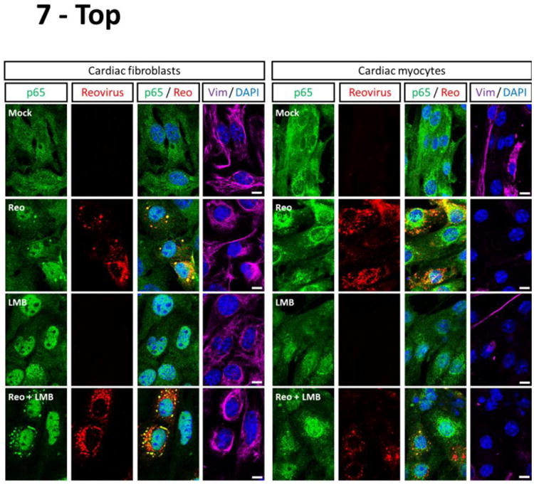
Primary cardiac cultures were infected with reovirus T3D or media alone (‘mock’). After incubation for 2 h, the inoculum was removed and replaced with either media alone or media containing 20 nM LMB. Cells were incubated for another 6 h prior to fixation and immunostained as indicated. The apparent co-localization of RelA within reovirus viral factories is due to cross-reactivity of the p65 antibody with a reovirus protein (data not shown). Scale bar = 10 μm. The percentage of cardiac fibroblasts (n = 8 – 104 cells per condition) or cardiac myocytes (n = 18 – 48 cells per condition) displaying nuclear p65 is indicated for a representative of two independent experiments. For cases that received reovirus inoculum, only cells positive for reovirus antigen were scored.