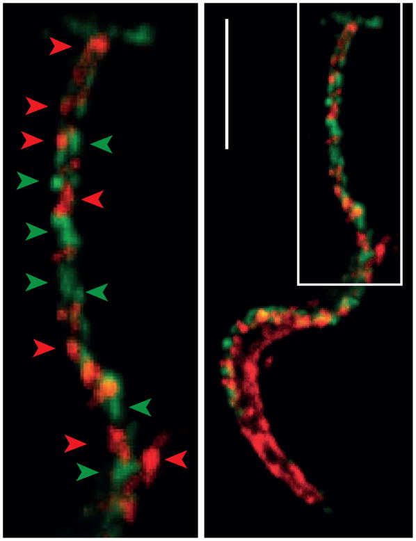
Figure 1. trans-Sialidase and Mucins Localize to Distinct Nanodomains on the Trypanosoma cruzi Trypomastigote Coat.
Upper: T. cruzi trypomastigote domains studied by confocal microscopy. Lower: Magnification of the flagellum. Arrowheads denote different membrane domains (in green, sialic acid revealed with anti-Flag antibody and in red trans-Sialidase revealed by anti-shed acute phase antigen (SAPA) antibody). Bar: 5 μm.

