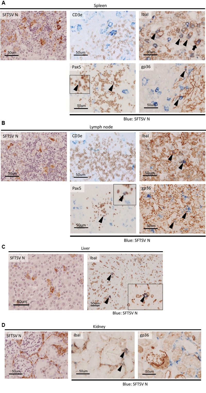
FIGURE 4.

Colocalization of host cell markers and SFTSV antigen in SFTSV-infected tissues from IFNAR-/- mice. The spleen (A) lymph node (B), liver (C), and kidney (D) of the SFTSV-infected mice were subjected to double staining with anti-SFTSV N antibodies and host cellular markers: CD3e (T cells), IbaI (macrophages), Pax5 (immature B cells), and gp36 (reticular cells). Cells stained with both SFTSV and host cellular markers are indicated by arrowheads, and the phagocytosis of an infected cell by an IbaI-positive macrophage is indicated by an arrow.
