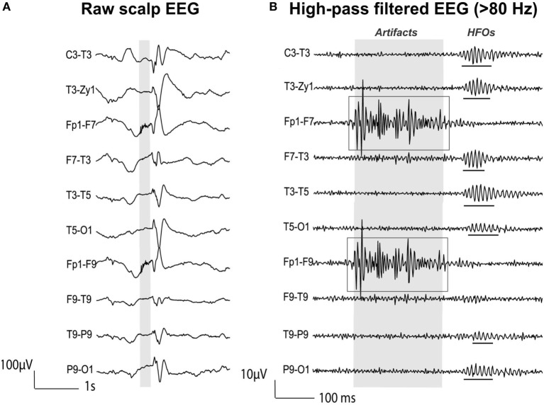
Figure 5.
High-frequency oscillations (HFOs) vs. artifactual oscillations recorded with scalp electroencephalography (EEG) data from a patient with focal epilepsy. (A) Raw EEG with interictal epileptic spikes (gray section). (B) EEG filtered with high-pass filter of 80 Hz. Gray section in (A) is expanded in time and amplitude in (B). Ripple HFOs are underlined. The morphology of HFOs is more rhythmic and regular in amplitude and frequency than artifactual oscillations. Source: adapted from Andrade-Valenca et al. (110).

