
Figure 3.
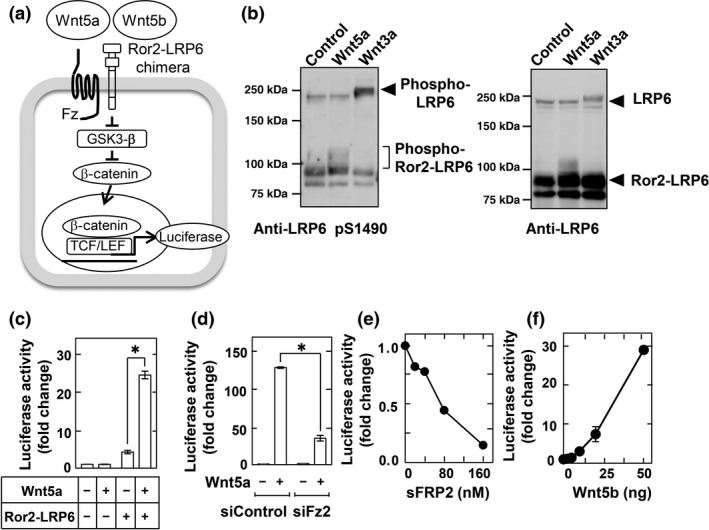
Development of reporter assay to measure Wnt5a and Wnt5b signaling. (a) Schema of the reporter system used to measure Wnt5a/b signaling using CHO cells stably expressing a Ror2– low‐density lipoprotein receptor‐related protein 6 (LRP6) chimera and TOP‐flash (CHO/Ror2‐LRP6‐TOP cells). GSK3‐β, glycogen synthase kinase 3β; TCF/LEF, T‐cell factor/lymphoid enhancer factor. (b) CHO/Ror2‐LRP6‐TOP cells were stimulated with 1 mL control, Wnt3a, or Wnt5a conditioned medium (CM) for 3 h, and cell lysates were probed with anti‐phospho‐LRP6 and anti‐LRP6 antibodies. (c) CHO/Ror2‐LRP6‐TOP cells and parental CHO cells stably expressing only TOP‐flash were stimulated with control or Wnt5a CM for 8 h. A luciferase assay was carried out and the activity was expressed as a fold increase compared with samples without Wnt5a and Ror2‐LRP6. Results are shown as means ± SD of three independent experiments. *P < 0.05. (d) CHO/Ror2‐LRP6‐TOP cells, which had been transfected with control or Frizzled‐2 (Fz2) siRNAs, were stimulated with control or Wnt5a CM for 8 h. (e) CHO/Ror2‐LRP6‐TOP cells were stimulated with Wnt5a CM for 8 h in the presence of the indicated concentrations of secreted Frizzled‐related protein‐2 sFRP2. (f) CHO/Ror2‐LRP6‐TOP cells were stimulated with the indicated concentrations of purified Wnt5b for 8 h.