
Figure 4.
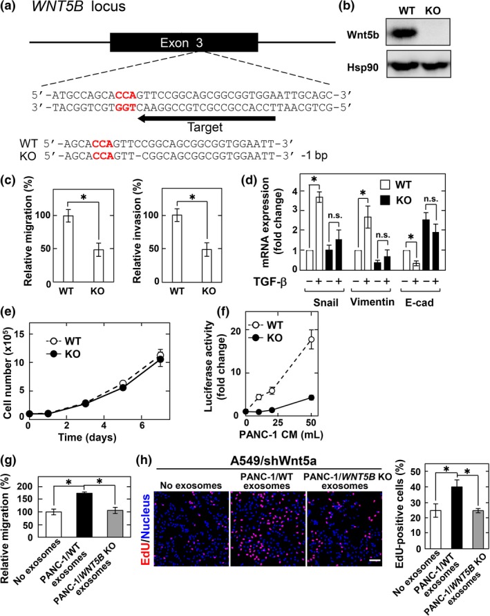
Wnt5b‐associated exosomes from PANC‐1 cells are active. (a) Schematic drawing of the targeting site of single guide RNA (sgRNA) at exon 3 of human WNT5B gene, and the nucleotide sequences of both alleles from wild‐type (WT) and WNT5B knockout (KO) PANC‐1 cells are shown. A dash represents a single base deletion. (b) Lysates from WT and WNT5B KO cells were probed with anti‐Wnt5b antibody. Hsp90, heat shock protein 90. (c) WT and WNT5B KO PANC‐1 cells were subjected to migration and invasion assays and relative migration and invasion activities of WNT5B KO PANC‐1 cells were expressed as percentages of those of wild‐type PANC‐1 cells. Results are shown as means ± SD of three independent experiments. *P < 0.05. (d) WT and WNT5B KO PANC‐1 cells were stimulated with 5 ng/mL transforming growth factor‐β (TGF‐β) for 48 h and mRNA levels of Snail, vimentin, and E‐cadherin (E‐cad) were measured by quantitative real‐time PCR. The results were expressed as fold‐changes compared with WT cells without TGF‐β stimulation. n.s., not significant. (e) WT and WNT5B KO PANC‐1 cells were subjected to cell proliferation assay. (f) CHO/Ror2‐LRP6‐TOP cells were treated with the P100 fraction recovered from the indicated volumes of PANC‐1 conditioned medium (CM) for 8 h. (g) A549/shWnt5a cells were subjected to migration assay in the presence of the exosomes isolated from CM of WT and WNT5B KO PANC‐1 cells and relative migration activity of A549/shWnt5a cells were expressed as percentages of those of A549/shWnt5a cells in the absence of the exosomes. (h) A549/shWnt5a cells were treated for 48 h with the exosomes isolated from CM of WT and WNT5B KO PANC‐1 cells, and then the cells were incubated with 5‐ethynyl‐2′‐deoxyuridine (EdU) for 1 h before fixation and stained with DRAQ5. EdU‐positive cells are expressed as percentages of positively stained cells compared with total DRAQ5 stained cells per field (n = 200–300). Scale bar = 100 μm.